What Is Histiocytoma in Dog, Symptoms, Causes and Treatment?

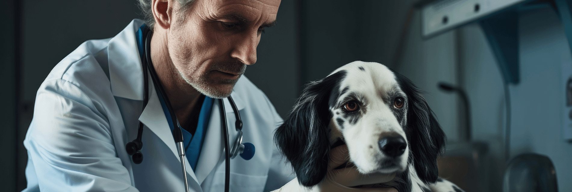
So, your dog has a strange lump or growth, and the vet says it’s histiocytoma. Don’t panic, it’s usually not a big deal. Histiocytomas are a type of skin growth in dogs that appears suddenly and just disappears on their own in a few months.
But you probably still have questions about what exactly they are, how to treat them, and if there’s any way to prevent them. This article will give you the lowdown on these common skin lumps, so you know what to expect.
We’ll go over what histiocytomas are, how they’re diagnosed and treated, how long they typically last, and when you should worry. You’ll be an expert on histiocytoma in dog in no time and feel more at ease about this sudden, small bump in the road. Read on to get the details about this temporary nuisance.
What is Histiocytoma in Dogs?

Histiocytoma in dogs is a benign skin tumor or growth that occurs most commonly in young dogs under 3 years of age. It is caused by the overgrowth of a type of immune cell called Langerhans cells in dogs. These cells are a part of a dog’s immune system and are present in the skin.
These cells play a crucial role in determining how a dog’s skin will respond to foreign material. The Langerhans are also responsible for the development of rashes when the dog’s skin is exposed to any irritants.
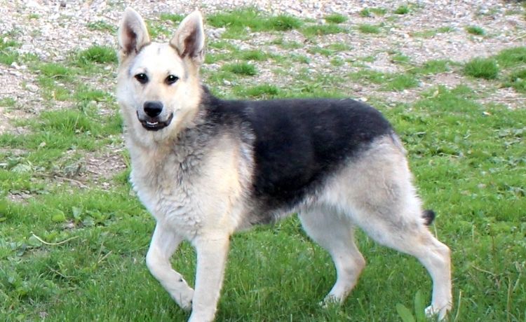
Also known as Cutaneous histiocytoma or button tumors, these skin growths usually appear as small, round, hairless, red or pink lumps on the head, ears, neck, or limbs. They may grow rapidly over a few weeks, but often regress spontaneously within a few months.
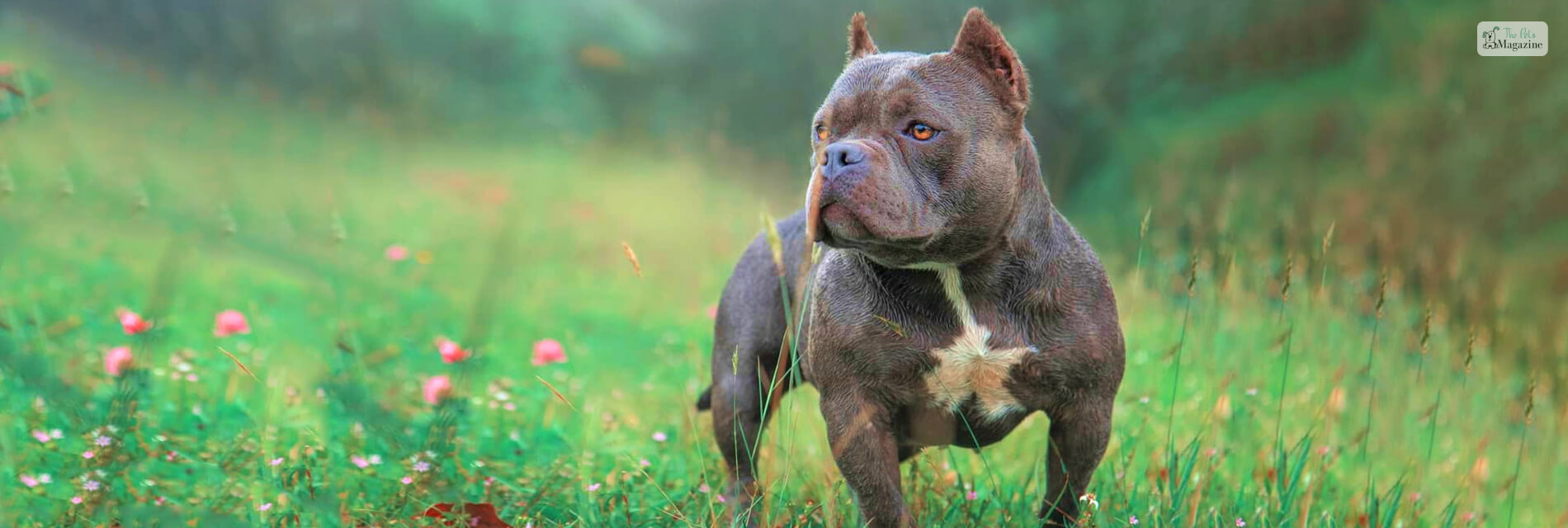
Histiocytoma can affect all dog breeds, including crossbreeds. However, certain breeds are more prone to histiocytoma. This includes Labrador Retriever, Boston Terriers, Dachshunds, Bulldogs, Scottish Terriers, American Pit Bull Terriers, Greyhounds, Shar Peis and Boxers.
Histiocytoma in dog is not usually painful or itchy, unless the growth becomes infected or irritated by the dog. They can be diagnosed by a fine needle aspirate or a biopsy of the mass. Moreover, Histiocytomas are not cancerous and do not spread to other parts of the body.
They are not contagious either and do not spread via skin to skin contact with other dogs. Furthermore, these types of skin growth do not pose any harm to humans or any other animals in the house, such as cats.
So, if you notice a new skin growth on your dog, don’t start panicking right away. Carefully monitor the mass for changes, and if it doesn’t disappear on its own in 4-6 weeks, you should talk to the vet.
Symptoms And Signs of Histiocytoma in Dog
In general, histiocytoma in dogs has no signs or symptoms. However, dogs develop a lot of lumps, bumps and cysts on their skin which says a lot about their overall health condition. Hence it is important for pet parents to know what a histiocytoma looks like. It will help them to understand what signs to watch out for and when to call the vet. Here are some symptoms and signs associated with histiocytomas in dogs:
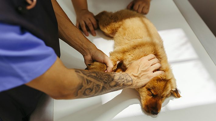
Skin Lump or Nodule
The primary sign of a histiocytoma is the development of a solitary, round, well-defined lump or nodule on the skin. These growths are usually small, ranging from a few millimeters to a few centimeters in diameter (usually less than 25 cm). They often have a button or dome-shaped appearance and can be pink, red, or flesh-colored. The surface of the nodule may or may not be ulcerated.
Rapid Growth
Another noticeable feature of histiocytoma in dogs is that these masses grow quickly over a few weeks. You may notice a sudden appearance and subsequent enlargement of the skin lump within a relatively short time frame. However, histiocytomas don’t appear overnight – they take around 1-4 weeks to develop.
Location
Histiocytomas most often appear on the head, ears, and legs of dogs. The hairless lumps tend to develop on areas with little fat and thin skin. While they can appear anywhere, the head and extremities are common sites.
Itching or Irritation
In some cases, histiocytomas can cause mild itching or irritation in dogs. You may observe your dog scratching, licking, or chewing at the site of the tumor due to discomfort.
Well-Demarcated Borders
Histiocytomas typically have well-defined, raised borders, meaning that you can easily distinguish the tumor from the surrounding healthy skin.
Self-Resolution
One characteristic feature of histiocytomas is their tendency to spontaneously resolve on their own. In most cases, histiocytomas will begin to shrink and disappear within a few months without any treatment. This spontaneous regression is more commonly seen in young dogs.
If you notice any of these symptoms in your dog, especially a fast-growing lump, schedule an appointment with your vet as soon as possible. They can perform a thorough physical examination, potentially conduct a fine-needle aspiration or biopsy, and provide an accurate diagnosis.
This is because other skin growth in dogs, including more serious tumors, can present with similar symptoms, so it’s always best to consult with a professional for proper evaluation and guidance.
Causes of Histiocytoma in Dog

While the exact cause of histiocytomas is not fully understood, there are several factors that may contribute to their development. Here are some possible causes and contributing factors:
Immune system dysregulation
It is believed that histiocytomas may develop because of an abnormal immune response. The immune system may mistakenly target certain cells in the skin, leading to the formation of the tumor.
Breed predisposition
Certain dog breeds, such as Labrador Retrievers, Boxers, and Staffordshire Bull Terriers, are more commonly affected by histiocytomas. This suggests that there may be a genetic component involved in their development.
Hormonal factors
Hormonal imbalances or fluctuations, particularly during puberty, have been suggested as a potential trigger for histiocytoma formation. This is supported by the fact that these tumors often regress spontaneously as the dog matures.
Viral or infectious triggers
Some research suggests that viral or infectious agents may play a role in the development of histiocytomas, although no specific pathogen has been definitively identified.
Environmental factors
Exposure to certain environmental factors, such as ultraviolet (UV) radiation, chemicals, or toxins, has been proposed as a potential cause. However, there is limited evidence to support this hypothesis.
It’s important to note that histiocytomas are generally benign and tend to resolve on their own without treatment. However, if you notice any unusual growths or skin abnormalities on your dog, it is always recommended to have them examined by a veterinarian for proper diagnosis and treatment.
Diagnosis and Treatment of Histiocytoma in Dog

To diagnose a histiocytoma in a dog, a veterinarian will typically perform a thorough physical examination and may need to conduct additional tests such as biopsy to verify the diagnosis.
So, you probably want to know what happens during the physical exam! We will tell you, so read on!
During the physical exam, your vet will thoroughly inspect the tumor. They will check the appearance and characteristics of the tumor to ensure that it meets the criteria – Histiocytomas are usually round, raised bumps that are pink to red in color. They tend to be firm and smooth, ranging from 0.5 to 2 inches in diameter.
Your vet may also order blood tests or imaging like x-rays to check for any underlying conditions or see if the growth has spread. However, one of the ways to correctly diagnose a histiocytoma is through a biopsy. There are mainly two types of biopsies – full excision biopsy and punch Biopsy.
Your vet will take a small sample of the growth to examine under a microscope. They will check for the presence of histiocytes, which are a type of white blood cell. An overabundance of these cells confirms the diagnosis of a histiocytoma.
Fine-needle aspiration (FNA) is another common method used to assess and determine histiocytoma in dogs. In this method, a needle is used to collect a sample of cells from the tumor. The cells are then examined under a microscope to confirm the diagnosis of a histiocytoma.
What Are the Next Steps After Diagnosis?
Once diagnosed, most histiocytomas do not require any treatment and will disappear on their own in a few weeks to months. However, in certain cases, the vet may suggest some alternative treatment options. For instance, some tumors may require surgery to remove them, especially if the growth is irritating your dog or is in a location where it could get injured.
Treatment of Histiocytoma in Dogs

Most histiocytomas in dogs regress spontaneously without treatment within a few weeks to a few months. However, some may persist or cause complications, in which case treatment options may include:
Waiting and Observing
The most conservative approach is to simply monitor the histiocytoma. If the histiocytoma is small and not causing any discomfort to the dog, the veterinarian may recommend monitoring it closely without immediate treatment. Take photos to track its size and appearance. If it’s not causing discomfort or bothering your dog in any way, it will likely shrink significantly or disappear completely within 2 to 3 months.
Steroid medication
Oral or topical steroids, such as prednisone or cortisone cream, may help speed up the regression of histiocytoma. This is because steroids work by reducing inflammation and the body’s immune response. Side effects of using steroids on dogs are usually minimal but may include increased thirst, appetite, and urination.
Surgical removal
If the histiocytoma is large, ulcerated, or in an area that is causing your dog discomfort or there is concern about its appearance, the veterinarian may recommend surgical excision. This procedure is relatively minor and involves removing the tumor under general anesthesia, light anesthesia or sedation. The histiocytoma is excised down to its base to remove it completely. After removal, the surgeon will use stitches or surgical glue to close the incision. Surgical removal is usually curative, and the tumor is unlikely to recur.
In the case of surgical removal of histiocytoma in dogs, the recovery process and precaution will be like that of any other minor canine surgery. You should not give your dog a bath for at least 2-3 weeks or until the incision completely heals. They should also not engage in any vigorous activities like zoomies around the yard, hiking or agility training.
Also make sure that your dog is not licking or scratching the incision or the surrounding area. You can put an e-collar or a cone on your dog to stop them from irritating the area around the incision.
Cryosurgery
Cryosurgery involves freezing the tumor with liquid nitrogen, which destroys the abnormal cells. Freezing the histiocytoma with liquid nitrogen destroys the cells, causing the bump to slough off in 7 to 14 days.
It is a non-invasive procedure and can be an alternative to surgery, particularly for smaller tumors. In case of large tumor, multiple treatment of cryosurgery may be required for it to be fully effective. But skin cutting can be avoided in this treatment method. Risks are minimal but may include light scarring or skin discoloration.
During cryosurgery treatment, the vet will use a cryoprobe to spray liquid nitrogen on the tumor. Cryoprobe is a small and insulated gadget used to disperse or circulate liquid nitrogen over the affected area and kill diseased tissue. The vet may also dab the liquid nitrogen on the tumor with a cotton or a foam swab. Before the cryosurgery procedure, the vet will use a local anesthetic to numb the surrounding area. So, you can rest assured because your dog will experience only minor discomfort.
Aftereffects of Cryosurgery in dogs
Cryosurgery, while minimally invasive, can cause some aftereffects in dogs. Here’s what to expect:
Short-term pain: The freezing process itself can be painful due to cellular disruption and nerve irritation. Vets will use anesthesia during the procedure to address this.
Swelling and redness: The treated area will likely swell and appear red for a few days after the procedure. This is normal inflammation as the body reacts to the dead tissue.
Sloughing and odor: Over the next 1-2 weeks, the dead tissue will dry up and fall off. This might look unsightly and even have a foul odor. It’s important not to pick at it and let it shed naturally.
Discomfort during shedding: While the nerves are damaged during freezing, there might be some discomfort as the dead tissue loosens and falls off.
Multiple treatments: Depending on the size and severity of the condition, your dog might need multiple cryotherapy sessions.
While these aftereffects might seem concerning, they are generally temporary and part of the healing process.
Recovery After Surgery
Here are some ways to help your dog recover after a minor surgery like Histiocytoma removal:
Create A Comfy Recovery Space
Before your dog gets back from the veterinary clinic or the hospital after undergoing surgery for histiocytoma, pick a quiet area in your house. Make this area as cozy and comfortable as possible so that your dog can rest and recover after surgery without disturbance. Provide a soft bed with enough space for them to stretch out comfortably. This will help prevent pressure on the incision site. Also place a soft blanket, pillows or cushions on the dog bed so that they have a soft and cozy place to tuck away and relax.
Limit Activity
After surgery, your dog will need to restrict their activity for a while, usually for about 7-14 days or until the sutures are removed. This includes all activities that can put a strain on the area of the incision such as running, jumping, or playing rough. Unless your vet has specifically suggested, you should also avoid taking your dog for a swim. Also consult your veterinarian about the recommended activity level during the recovery period and duration of restriction.
Monitor The Incision Site
Keep a close eye on the incision for any signs of redness, swelling, or discharge. These could be signs of infection. Make sure that your dog is not licking or chewing the incision which can slow healing and increase the risk of infection. If they are, then you will have to make your dog wear a Elizabethan collar (also known as pet cones) to stop from licking or chewing and further irritating the incision area.
Manage Pain And Medication
Your veterinarian may prescribe pain medication for your dog to help with post-op pain and discomfort. Follow the vet’s instructions carefully regarding dosage and frequency. If you have a dog who is high-strung or gets anxious easily, then inform your vet beforehand and they may prescribe a sedative or anti-anxiety medication.
Food And Hydration
Your dog might lose their appetite after surgery due to anesthesia. Offer small, bland meals like cooked chicken and rice initially. Gradually reintroduce their regular food as their appetite returns. Also ensure your dog has access to fresh water at all times.
Follow-Up Care
Schedule a follow-up appointment with your veterinarian to check on the healing progress and remove any stitches or bandages if needed.
Final Thoughts
So, by now you probably have a good idea of what histiocytoma is and what to expect if your dog develops one. The good news is that these growths are typically benign and will often disappear on their own in a few weeks or months. While waiting for it to regress, keep an eye on it to ensure it’s not changing or getting bigger, and have your vet examine it if you notice any concerning changes.
However, if it doesn’t regress after a few months, then you should consult your vet and follow their guidance regarding the diagnosis and treatment of your dog’s histiocytoma. They will be able to provide the most accurate assessment and recommend the most appropriate course of action based on the specific circumstances of your dog’s condition.
You May Also Like..
Share


All Comments
Corbin Haley
4 March, 2024
You always manage to deliver quality content that's both informative and engaging.
Reply